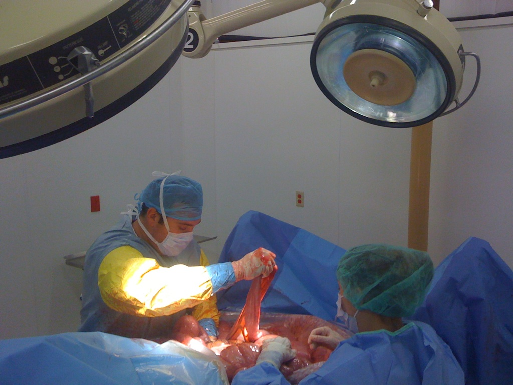
CALEC surgery, or Cultivated Autologous Limbal Epithelial Cell surgery, offers groundbreaking hope for individuals suffering from corneal damage previously considered irreparable. This innovative procedure harnesses the potential of stem cell therapy, utilizing limbal epithelial cells harvested from a healthy eye to regenerate the damaged corneal surface. Clinical trials led by Ula Jurkunas at Mass Eye and Ear have demonstrated CALEC’s impressive effectiveness, achieving over 90% success rates in restoring vision and alleviating pain in patients affected by eye injuries. As a revolutionary alternative to traditional corneal transplant methods, CALEC surgery significantly improves the prospects for those facing limbal stem cell deficiency. With a promising safety profile and remarkable outcomes, this surgery may transform the landscape of ophthalmic treatments, fostering hope for countless individuals worldwide.
Cultivated autologous limbal epithelial cell (CALEC) procedures represent a novel advancement in addressing corneal impairments resulting from severe ocular injuries or diseases. By leveraging regenerative medicine techniques, specifically stem cell therapies, these surgical interventions focus on restoring the ocular surface using epithelial cells derived from a patient’s own eye. Patients suffering from limbal epithelial cell deficiency now have a groundbreaking alternative to conventional corneal transplants, with the potential for long-term corneal surface restoration. The clinical application of CALEC reflects a significant stride towards not only repairing eye damage but also enhancing visual acuity for individuals who previously had limited treatment options. As researchers continue to optimize these procedures, the hope of broader applications and improved outcomes for eye health remains a primary goal.
Understanding CALEC Surgery and Its Significance
CALEC surgery, or cultivated autologous limbal epithelial cell surgery, marks a groundbreaking advancement in the field of ophthalmology. This innovative procedure focuses on harnessing the regenerative abilities of limbal epithelial cells, which are crucial for maintaining the cornea’s surface integrity. By using stem cells from a healthy eye, surgeons can cultivate a cellular graft that has shown remarkable success rates in clinical trials. This surgical approach is particularly important for patients suffering from severe corneal damage due to trauma, chemical burns, or infections, where conventional treatments, such as corneal transplants, may not be viable.
The significance of CALEC surgery lies not only in its technical capability to restore vision but also in the hope it offers to patients who once faced bleak prognoses. As reported in recent clinical trials, CALEC has achieved over 90 percent effectiveness in regenerating the corneal surface, thus providing a new lease on life for those affected by persistent ocular surface disorders. The procedure’s design allows for the continued use of the patient’s own tissues, minimizing rejection risks and complications commonly associated with donor grafts.
The Role of Stem Cell Therapy in Eye Injury Repair
Stem cell therapy represents a revolutionary advancement in the landscape of regenerative medicine, providing innovative solutions for repairing ocular injuries that were previously deemed irreversible. The use of limbal epithelial cells derived from stem cells has opened new avenues for treating corneal surface damage. These stem cells are integral to maintaining the health and function of the eye, allowing for effective restoration of the corneal surface after sustaining injuries. In the context of CALEC surgery, stem cell therapy has proven to be a safe and effective means to rejuvenate the corneal surface and improve visual outcomes.
The success of stem cell therapy in the context of CALEC surgery highlights the potential for broader applications in treating various eye injuries and conditions. The retrieval and cultivation of limbal epithelial cells from a healthy eye provide a personalized approach that helps avoid complications related to foreign tissue rejection. By rehabilitating the cornea with the patient’s own cells, healthcare providers can significantly reduce the instance of postoperative complications, thereby enhancing patient safety and satisfaction.
Benefits of Limbal Epithelial Cell Cultivation
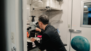
The cultivation of limbal epithelial cells is a critical component of CALEC surgery, enabling surgeons to generate a biological graft that promotes corneal restoration. This innovative process has evolved through years of research and clinical trials, validating its safety and effectiveness. By expanding a small number of limbal epithelial cells harvested from the healthy eye, this technique not only facilitates the regeneration of corneal surface layers but also helps restore visual function and alleviate the persistent pain associated with corneal injuries.
Furthermore, the advancement in technologies surrounding cell cultivation ensures that the grafts meet stringent quality and safety standards, thereby enhancing the overall success rates of the procedure. This meticulous approach aids in creating a sustainable source of limbal epithelial cells, paving the way for additional clinical applications. As further research progresses, the potential to adapt this technique for treating bilateral eye damage holds promise for widening the scope of patients who can benefit from CALEC surgery.
Challenges and Future Directions in CALEC Clinical Trials
Despite the significant success achieved with CALEC surgery in early clinical trials, several challenges remain that need to be addressed for broader implementation. One primary concern is the requirement that patients only have one affected eye to provide necessary material for the cell graft. Future research endeavors aim to establish an allogeneic approach that would utilize limbal stem cells from donor eyes, thereby expanding accessibility for individuals with damage to both eyes.
Moreover, as CALEC surgery is still considered experimental, ongoing studies will focus on larger patient cohorts and multi-institutional collaborations to further validate the efficacy and safety of the procedure. These efforts are essential for gathering sufficient data to support FDA approval and to ultimately enhance the availability of this promising treatment option for eye injuries. Researchers and clinicians remain optimistic about the potential for CALEC surgery to transform the treatment landscape for corneal disorders.
The Impact of CALEC on Visual Acuity Restoration
The impact of CALEC surgery extends beyond mere corneal surface restoration; it also significantly influences visual acuity among patients suffering from severe ocular injuries. Clinical trials have demonstrated varying degrees of improvement in visual function, with notable increases in the quality of life for participants. Patients who previously endured debilitating visual impairments reported enhanced clarity of sight, highlighting the profound implications of successful CALEC treatment.
Additionally, the assessment of visual outcomes following CALEC surgery underscores the importance of this therapeutic approach in addressing chronic vision loss. With promising results indicating a restoration of vision for a significant percentage of trial participants, the future of stem cell-derived treatments in restoring sight appears bright. This successful restoration can allow individuals to reclaim their independence and improve their daily functioning, thereby highlighting the societal impact of innovative ocular treatments.
Innovations in Corneal Surface Restoration Techniques
Innovative techniques in corneal surface restoration continue to evolve, driven by advancements in stem cell therapy and regenerative medicine. CALEC surgery stands at the forefront of these innovations, utilizing a highly specialized method to cultivate individual cells that promote healing and recovery. The procedural framework not only incorporates advanced surgical techniques but also leverages state-of-the-art manufacturing processes to ensure the viability and effectiveness of the grafts used in treatment.
These advancements are paving the way for future research that aims to refine existing methodologies while exploring new avenues for cellular therapy. As the field of regenerative ophthalmology progresses, it is anticipated that further innovations will emerge, enhancing the ability to treat diverse ocular conditions effectively. Researchers are investigating the integration of biomaterials and tissue engineering, which may expand the applications of corneal surface restoration techniques even further.
Preparing Patients for CALEC Surgery
Preparing patients for CALEC surgery involves a thorough preoperative assessment and education regarding the procedure’s benefits and limitations. Potential candidates must undergo a comprehensive evaluation to ensure they meet the criteria for the procedure, particularly focusing on the health of their unaffected eye, which is essential for obtaining limbal epithelial cells. Informed consent is another crucial aspect of the preparation process, ensuring that patients understand the procedural risks and expected outcomes.
Additionally, healthcare providers emphasize the importance of post-surgical care to maximize recovery and visual outcomes. Patients are often educated on behaviors and precautions necessary for promoting successful healing of the cornea post-operation. This holistic approach to patient management not only prepares individuals for the surgical process but also fosters positive ongoing relationships between patients and their healthcare teams.
Regulatory Aspects of CALEC and Future Trials
As CALEC surgery transitions from experimental trials toward possible standardization in clinical practice, navigating regulatory aspects becomes increasingly important. The oversight by bodies such as the U.S. Food and Drug Administration ensures that therapies meet required safety and efficacy benchmarks before they can be widely implemented. Clinical trials thus play a pivotal role in collecting necessary data that will ultimately inform regulatory decisions.
Future trial designs are likely to include larger patient populations and randomized control groups, strengthening the credibility of findings and expediting the path to approval. Collaboration with regulatory authorities will be essential in ensuring that CALEC procedures can provide effective treatment for a more extensive range of patients suffering from corneal injuries.
Advancing the Understanding of Limbal Stem Cell Deficiency
Limbal stem cell deficiency represents a critical barrier to the effective treatment of various ocular surface disorders. Understanding the mechanisms that lead to this deficiency is vital for developing targeted therapies like CALEC. This condition arises when the limbal epithelial cells are depleted due to traumatic injury, chemical burns, or other insults to the eye. The ability to replenish these cells through stem cell therapy highlights the importance of ongoing research aimed at comprehensively understanding the factors contributing to limbal stem cell depletion.
Additionally, advancing knowledge in this area will not only bolster the development of effective treatment options such as CALEC surgery but also enhance preventive measures. The more that researchers uncover the pathophysiology surrounding limbal stem cell deficiency, the better equipped they will be to provide holistic approaches aimed at both treatment and prevention of corneal damage.
Frequently Asked Questions
What is CALEC surgery and how does it work?
CALEC surgery, or Cultivated Autologous Limbal Epithelial Cell surgery, is a groundbreaking procedure that involves taking limbal epithelial cells from a healthy eye and cultivating them to create a graft. This graft is then transplanted into a damaged eye to restore the corneal surface, revolutionizing treatment options for patients with severe corneal injuries.
What are the benefits of CALEC surgery for eye injuries?
CALEC surgery offers significant benefits for individuals with eye injuries by providing a safe and effective means of restoring the corneal surface. The procedure has demonstrated over 90 percent effectiveness in clinical trials, allowing patients with previously untreatable corneal damage to regain vision and alleviate pain.
How are limbal epithelial cells utilized in CALEC surgery?
In CALEC surgery, limbal epithelial cells are harvested from a healthy eye through a biopsy. These cells are then expanded to create a tissue graft, which is surgically implanted in the damaged eye, promoting restoration of the corneal surface that is essential for clear vision.
What types of corneal injuries can CALEC surgery help treat?
CALEC surgery can effectively address various types of corneal injuries, including those resulting from chemical burns, infections, or traumatic events leading to limbal stem cell deficiency. By restoring the corneal surface, it alleviates the associated pain and facilitates visual rehabilitation.
How does CALEC surgery compare to traditional corneal transplant procedures?
Unlike traditional corneal transplants, which may not be suitable for patients with limbal stem cell deficiency, CALEC surgery utilizes the patient’s own limbal epithelial cells to create a graft, making it a more viable option for restoring corneal surface integrity in difficult cases.
Is CALEC surgery currently available for patients?
As of now, CALEC surgery remains experimental and is not widely available in hospitals. However, clinical trials have shown promising results, and further studies are needed before the procedure can be offered as an approved treatment for eye injuries.
What are the success rates for CALEC surgery after the procedure?
In clinical trials, CALEC surgery showed success rates of 93 percent at 12 months and 92 percent at 18 months, with many patients experiencing significant improvements in visual acuity, indicating that the procedure effectively restores the corneal surface over time.
What safety measures are in place during CALEC surgery?
CALEC surgery has been shown to have a high safety profile, with no serious adverse events reported in study participants. Minor complications, such as infections, were promptly addressed, ensuring patient safety during the transplantation process.
Will CALEC surgery be available for patients with damage in both eyes?
Currently, CALEC surgery is limited to patients with damage in only one eye due to the need for a biopsy from the healthy eye. Future research aims to develop an allogeneic process using limbal stem cells from donor cadaveric eyes to expand eligibility.
What are the future prospects for CALEC surgery and clinical trials?
Future CALEC studies are aimed at enrolling larger patient populations at multiple centers, with longer follow-ups to further validate its efficacy and safety. Researchers hope that successful outcomes will lead to FDA approval and broader access to this innovative treatment for corneal injuries.
| Key Point | Details |
|---|---|
| Introduction of CALEC Surgery | First CALEC surgery performed at Mass Eye and Ear by Ula Jurkunas. |
| Clinical Trial | A clinical trial involved 14 patients and showed that CALEC effectively restored the cornea’s surface. |
| Procedure Description | Involves harvesting stem cells from a healthy eye, expanding into a graft, and transplanting into the damaged eye. |
| Effectiveness | 90% success rate in restoring corneal surfaces after 18 months. |
| Safety Profile | The procedure showed a high safety profile with no serious adverse events. |
| Future Research | Future studies planned to include larger patient groups and aim for FDA approval. |
Summary
CALEC surgery represents a groundbreaking approach in ocular medicine, providing new hope for patients with previously untreatable corneal damage. This innovative treatment, involving stem cell therapy, showcases a strong safety profile and high effectiveness rates, particularly in restoring the cornea’s surface. As the clinical trials progress and expand, there is an optimistic outlook that CALEC surgery will soon be accessible to a broader patient population, ultimately enhancing vision rehabilitation efforts.